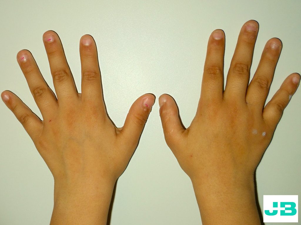
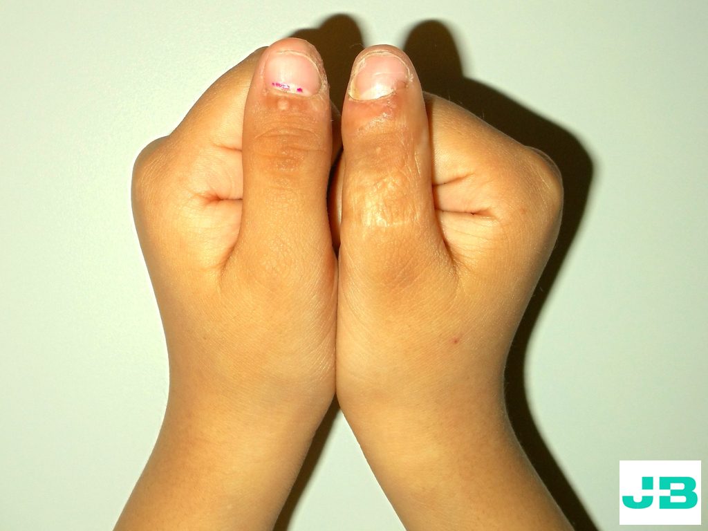
Clinical cases

Real cases explained in plain language. Some surgical images may be upsetting: they are blurred until you choose to view them.

Case 1

Osteoarthritis of the finger joints

Finger osteoarthritis is the most common degenerative condition of the hand and mainly affects the finger joints and the base of the thumb. It causes deformity and pain of varying intensity; in women, the deformity may be the main reason for consultation.

Case

A 56-year-old woman consulted for pain and deformity of the left index and middle fingers. Pain in the middle finger prevented her from making a full fist, reducing her grip strength. As physiotherapy and analgesics had not helped, joint replacement with a prosthesis was indicated for the middle finger, and arthrodesis (joint fusion) for the index finger.
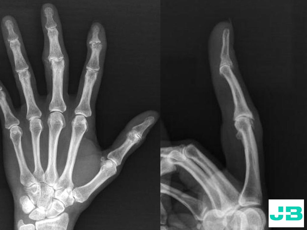
X-rays show the deformity of the index and middle finger joints.
FIG. 1. X-rays show the deformity of the index and middle finger joints.
VIDEO. The pre-operative video shows the deformity and lack of motion of the middle finger.
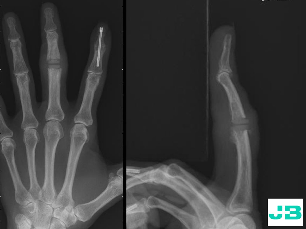
Post-operative X-rays: joint replacement of the middle finger and fusion of the index finger.
FIG. 2. Post-operative X-rays: joint replacement of the middle finger and fusion of the index finger.
VIDEO. After surgery, the patient regained a full, pain-free fist.
Case 2

Distal radius malunion and thumb flexor tendon rupture

Wrist fractures are the most common bone injuries of the upper limb. Although the best treatment is debated, especially in patients over 65, inadequate treatment can lead to sequelae such as pain, deformity and loss of function.

Case

A 61-year-old woman consulted three months after surgery for a left wrist fracture. She had deformity and limited wrist motion and, in addition, could not bend her thumb.

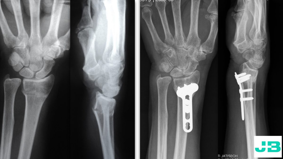
Left: the fracture. Right: the treatment performed, showing the fracture was not corrected. The plate, standing off the bone, injured the flexor tendons.
FIG. 1. Left: the fracture. Right: the treatment performed, showing the fracture was not corrected. The plate, standing off the bone, injured the flexor tendons.
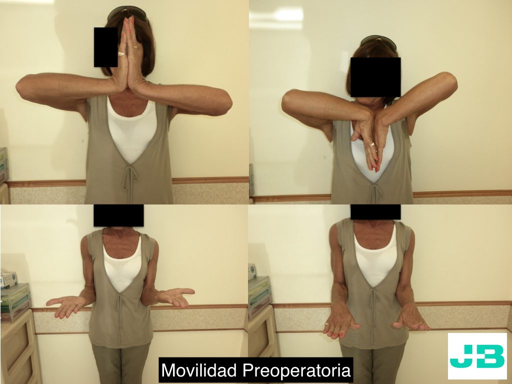
Pre-operative assessment: 30% loss of wrist flexion and extension compared with the healthy side, and pain on forearm rotation.
FIG. 2. Pre-operative assessment: 30% loss of wrist flexion and extension compared with the healthy side, and pain on forearm rotation.
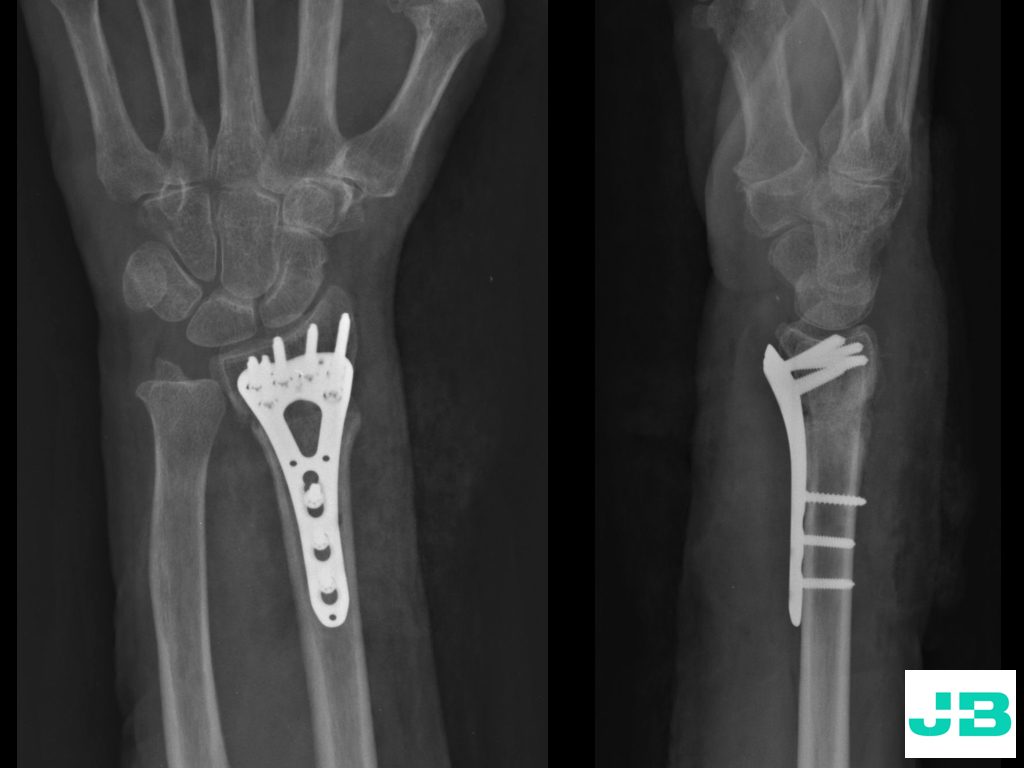
Post-operative X-rays: the deformity was corrected through an osteotomy and fixed with a plate and screws so the bone heals in good position.
FIG. 3. Post-operative X-rays: the deformity was corrected through an osteotomy and fixed with a plate and screws so the bone heals in good position.
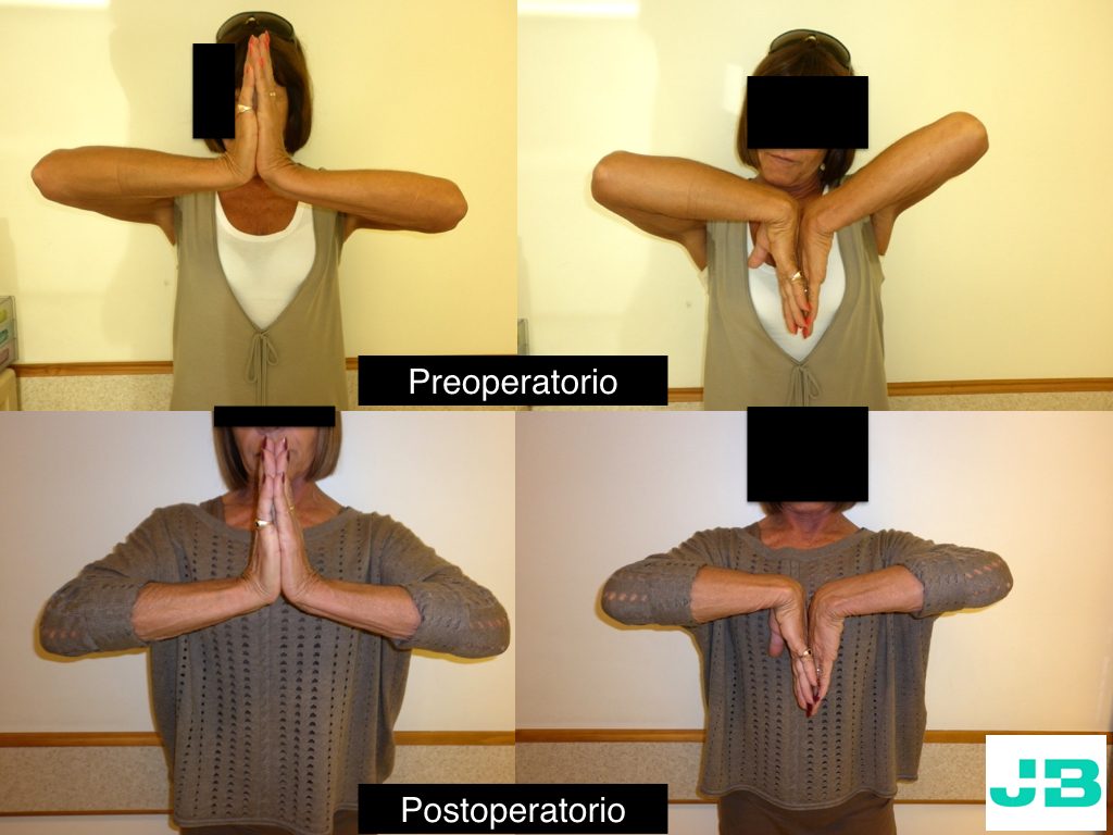
Recovery of motion: top images before surgery, bottom images after.
FIG. 4. Recovery of motion: top images before surgery, bottom images after.
VIDEO. Recovery of thumb flexion after reconstructing the flexor tendon with a graft.
Case 3

Radial nerve palsy

Radial nerve injury paralyses the muscles that extend the wrist, thumb and fingers, making it hard to pick up objects and weakening grip. The aim is to repair the nerve; when function cannot be recovered that way, a tendon transfer allows most upper-limb palsies to be reconstructed.

Case

A 33-year-old man consulted for a radial palsy of 5 years’ duration following a surgically treated humerus fracture. He underwent surgery to reconstruct finger and thumb extension through tendon transfers.

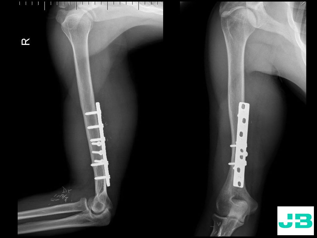
X-rays showing the earlier treatment of the humerus fracture.
FIG. 1. X-rays showing the earlier treatment of the humerus fracture.
VIDEO. Before surgery: no extension of fingers and thumb, with wrist extension preserved.
VIDEO. Final result: the patient regained finger and thumb extension, improving hand function.

Every patient is different: results may vary. Images are shown for information only.